Serena Williams once said, “I really think a champion is defined not by their wins but by how they can recover when they fall.”
Recovery is just as much a mental mindset as it is physical. This is why, for many athletes, learning about their bodies and finding tactics to facilitate recovery is where mental and physical mindsets converge.
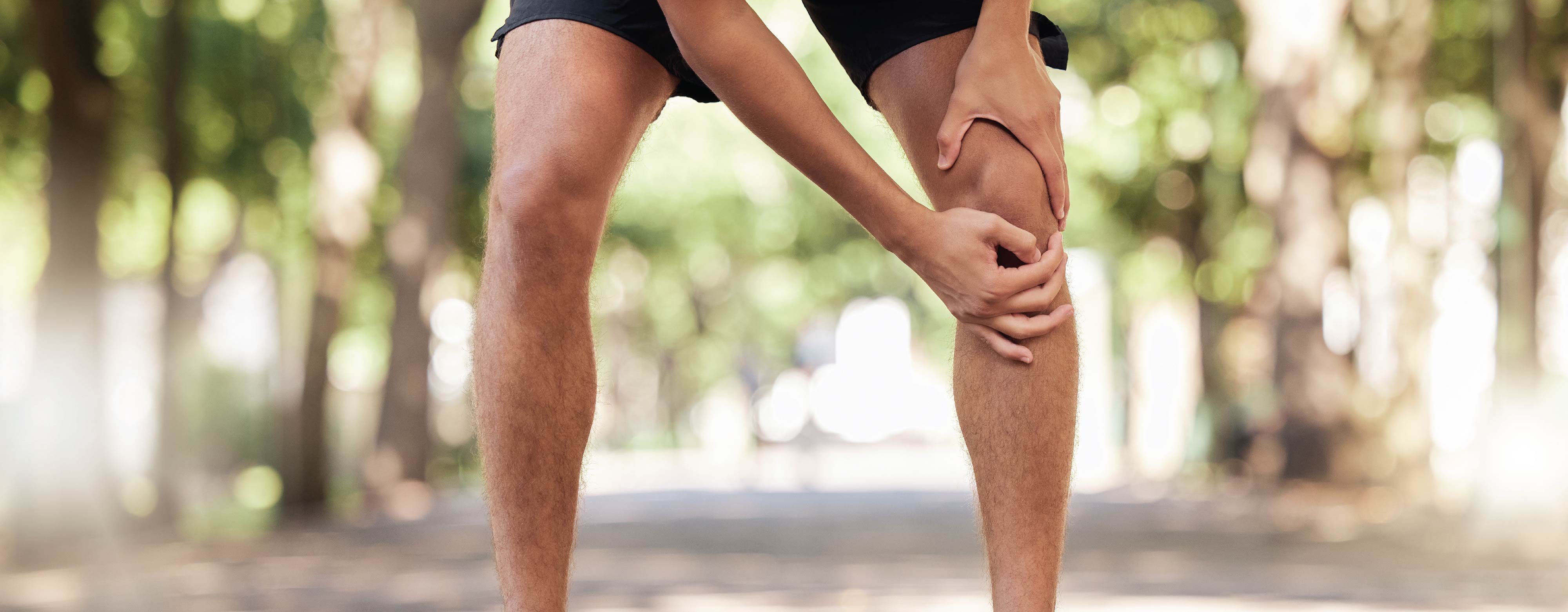
As with other high-intensity sports like basketball and soccer, tennis involves quick starts and stops, lateral movements, jumping and pivoting - over and over again. This means the knees take quite the beating whether you are elite or amateur.
The knees withstand up to 2-3 times our body weight when walking and 7-12 times our body weight when running (Herman, 2016; Taylor, Heller, Bergmann, & Duda, 2004). More than that, there are many moving parts in the knee joint, so if we suddenly move, step or jump in the wrong way, damage and pain can easily follow.
Indeed, it boils down to the neuromuscular coordination of our movements and our sense of awareness, which have the most influence over sustaining a knee injury. Typically, knee injuries are non-contact injuries.
The knee joint is as complex as the source of knee pain
For athletes, the knee joint is more than a hinge; it's a powerhouse for performance. Coordination is crucial, and loading capacity is critical. To start, four ligaments are arranged in a crisscrossing formation to ensure stability around the knee joint when we perform lateral movements, jump, stop and pivot. These include the:
1. Anterior Cruciate Ligament (ACL)
2. Posterior Cruciate Ligament (PCL)
3. Medial Collateral Ligament (MCL)
4. Lateral Collateral Ligament (LCL)

To absorb the intensity of these movements and counterbalance our body weight, two menisci (crescent-shaped pieces of cartilage) cushion the joint. The patellar tendon ensures forces are transmitted from our quadriceps to the tibia (shin bone) and the knee cap is aligned correctly during dynamic movements.
For example, all of these structures, ligaments, menisci, patella, and tendons are possible sources of pain. Many are enriched with nerve fibres responsible for signalling damage, initiating inflammatory responses, and thus eliciting pain.
One popular notion for more diffuse pain felt around or under the patella (knee cap), known as anterior knee pain or patellofemoral pain (PFP), is from misalignment or significant overload. It is one of the most common knee disorders experienced by athletes and accounts for about 30% of all injuries seen in sports clinics (LaBella, 2004).
While knee pain may be common, some athletes seek a colourful taping technique for their knees to bounce back sooner rather than later.
Kinesiology taping of quadriceps helps knee pain and muscle strength

Recovering from patellofemoral pain (PFP) can be challenging for athletes as the source of knee pain can come from several structures (Bolgla & Boling, 2011), including the underlying fat pad behind the patella. However, kinesiology tape has been proposed to improve lower-extremity neuromuscular control and thus may help to improve misalignment or overload issues of the knee joint.
In a recent study, kinesiology tape application over vastus medialis obliques (VMO), a quadricep muscle, in athletes with PFP improved pain and functional performance. In addition, gains in muscle strength were observed in the athletes following the exercise therapy during which kinesiology tape was worn (Aghapour, Kamali, & Sinaei, 2017).
It is, however, unclear if the improved (isokinetic) muscle strength results from pain relief induced by kinesiology taping or altered biomechanics. It is well known that quadriceps muscle weakness, especially for eccentric contractions, is usually present in persons with anterior knee pain.
Regardless, the addition of kinesiology taping comes without any risks. A recent review underscores that pain relief is the most common outcome where the authors write, "...the reduction in loading on the patellofemoral joint is a key point of positive influence" (Dehghan, Fouladi, & Martin, 2023).
Other alternatives to kinesiology taping for anterior knee pain
Athletes with anterior knee pain or PFP may choose other adjuvant therapies alongside their rehabilitation and training, such as:
• electrotherapy
• stretching and strengthening exercises
• balance training
• patellar braces
• patellar mobilization
• biofeedback training
 |
 |
 |
 |
As Serena Williams alluded, winning is about finding out how you get back into the game. Regarding kinesiology taping, there are no restrictions, as the tape can be worn during rehabilitation activities, training and matches. So when mental and physical mindsets converge, kinesiology tape emerges as an attractive and economical choice to wear on and off the court.
References
-
Aghapour, E., Kamali, F., & Sinaei, E. (2017). Effects of kinesio taping® on knee function and pain in athletes with patellofemoral pain syndrome. Journal of Bodywork and Movement Therapies, 21(4), 835-839. doi:10.1016/j.jbmt.2017.01.012
-
Bolgla, L. A., & Boling, M. C. (2011). An update for the conservative management of patellofemoral pain syndrome: A systematic review of the literature from 2000 to 2010. International Journal of Sports Physical Therapy, 6(2), 112-125. PMID: 21713229
-
Dehghan, F., Fouladi, R., & Martin, J. (2023). Kinesio taping in sports: A scoping review. Journal of Bodywork and Movement Therapies, doi:10.1016/j.jbmt.2023.05.008
-
Herman, I. P. (2016). Physics of the human body Springer.
-
Honore, Pretty (2022). "I'm Just Serena": Quotes From the GOAT That Will Keep You Motivated, https://www.distractify.com/p/serena-williams-quotes, Date of last access: September 18th, 2023.
-
LaBella, C. (2004). Patellofemoral pain syndrome: Evaluation and treatment. Primary Care: Clinics in Office Practice, 31(4), 977-1003. doi:10.1016/j.pop.2004.07.006
-
Taylor, W. R., Heller, M. O., Bergmann, G., & Duda, G. N. (2004). Tibio-femoral loading during human gait and stair climbing. Journal of Orthopaedic Research: Official Publication of the Orthopaedic Research Society, 22(3), 625-632. doi:10.1016/j.orthres.2003.09.003


Share:
How the most common shoulder injuries occur in overhead sports